13 Dec 2013

Two Ph.D. students – Mgr. Lucie Nováková and Mgr. Kristína Kovačovicová from the lab of MVDr. Martin Anger, Ph.D. (CEITEC Veterinary Institute Brno, Czech Republic) were successful in Leica Microsystem calendar contest. Their images are presented in Leica calendar 2014 – November and December.

A whole calendar is available at: Leica Microsystems Calendar 2014 – Now and Then

Standard 3D scanning modes in confocal laser scanning microscopy.

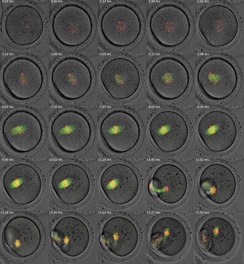
Meiotic Maturation: The image is a montage of selected time frames from a time-lapse recording of the meiotic maturation of a mouse oocyte. The images are an overlay of fluorescence images with images acquired using transmitted light as contrasting method.
 Polar Body Extrusion: Montage of spindles from mouse oocytes during the polar body extrusion process.
Polar Body Extrusion: Montage of spindles from mouse oocytes during the polar body extrusion process.
11 Dec 2013

Description from Nature Medicine, Dec 2013, Vol. 19, No. 12:
“Improved methods for identifying and tracking single podocytes in the intact kidney could improve our understanding of the mechanisms of glomerular injury and regeneration. In this issue (p1661), Janos Peti-Peterdi and his colleagues describe the use of Podocin-Confetti mice to image podocytes in vivo. The cover shows podocytes labeled in one of four colors (blue, green, yellow or red) and the renal vasculature.”

28 Sep 2013

Optical imaging devices have a finite depth of field and diffraction-limited resolution. The depth of field problem was tackled first with confocal microscopes; diffraction-unlimited resolution has been available for a few years now with super-resolution microscopes. Super-resolution microscopes with a solved depth of field problem are now available. Localization super-resolution uses astigmatism-based z-coding. STED super-resolution employs a mixed phase mask concept for tunable super-resolution in lateral and axial dimensions. Dr. Rolf T. Borlinghaus

Read more at: Super-resolution Microscopy and the Third Dimension